Cancer-busting particle beams have been caught in the act.
Particle beams can provide a blast of destructive energy directly to tumors — assuming the beam is in the right place. Now, using a radioactive beam, scientists pinpointed the beam’s location while treating tumors in mice. It’s the first successful treatment of tumors with a radioactive beam, scientists report in a paper submitted September 23 at arXiv.org.
The technique could eventually allow scientists to treat human patients with millimeter precision — particularly important when a tumor is nestled next to a sensitive organ such as the spinal cord or brain stem.
Various types of radiation can treat cancer. The most common is X-rays, high-energy light that can destroy the DNA in tumor cells. But X-rays deposit their energy all along the path of the beam, resulting in potential collateral damage in other parts of the body. More precise tumor targeting is possible with particles such as protons or ions — electrically charged atoms — which dump most of their energy in one spot.
Ion treatment is currently performed at more than a dozen centers worldwide. Those treatments use stable, non-radioactive ions — typically carbon-12, a variety of carbon with six protons and six neutrons in its nucleus. The particles in the beam have their electrons stripped off, giving them a positive charge.
The tumor is targeted based on calculations of how deep a beam will penetrate, coupled with previous imaging of the patient, for example, a CT scan (SN: 12/10/21). But bodies are not rigid, and organs can shift between imaging and treatment. Ideally, the beam’s position would be confirmed in real time. That’s just what the new technique allows.
“If you use a radioactive ion, you can simultaneously kill the tumor and see the beam,” says physicist Marco Durante of GSI Helmholtz Centre for Heavy Ion Research in Darmstadt, Germany.
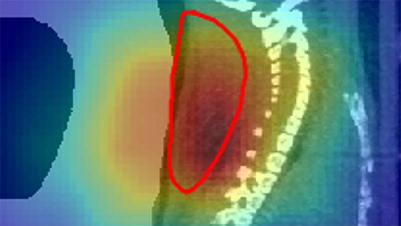
Durante and colleagues used carbon-11 ions, which have one fewer neutron in their atomic nuclei than carbon-12 ions do, making them radioactive. When carbon-11 decays, it releases a positron — a positively charged antimatter partner of an electron. Scientists can detect that positron annihilating with an electron in the body, via positron emission tomography, or PET (SN: 2/13/14). That identifies where the beam dumps its particles.
In the study, scientists used the carbon-11 ions to treat mice with tumors near the spine. Scientists were able to check the position of the beam during treatment and confirm that it was spot-on. Sure enough, the treatment shrunk the tumors.
Scientists had already tried to use PET to measure the location of a beam of stable ions. The stable ions don’t emit positrons, but some of the stable atomic nuclei break apart as they pass through material. Those fragments can make radioactive ions that release positrons in their decays. But the technique is difficult as the number of such particles is small.
With radioactive ion beams, many more positrons are emitted. “That allows [you] to get a very crisp and beautiful image of where the particle stops,” says radiation physicist Mitra Safavi-Naeini of Australia’s Nuclear Science and Technology Organisation in Sydney, who was not involved with the research.
The technique could also help scientists understand how radioactive material moves through the body after an ion treatment, Safavi-Naeini says. The radioactive particles are washed out of the beam’s bull’s-eye by blood flowing through the body. This spreads out the positron signal over time. The amount of this washout could help scientists understand if blood vessels are being destroyed by the treatment, thereby cutting off the tumor’s energy supply. This could help scientists understand how best to use particle beams to ensure the cancer’s demise.